Using inorganic dyes to label cells allows imaging by fluorescence and resonance Raman spectroscopy at the same time, which could help understand cell death caused by cancer and strokes say scientists in Ireland.
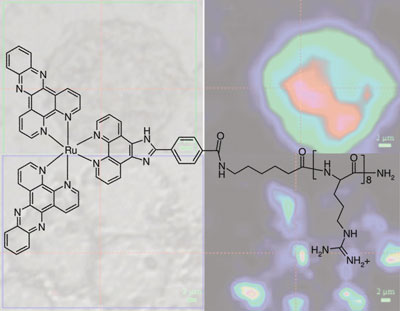
Fluorescence imaging and Raman mapping are two common techniques used to study live cells and understand the processes occurring in diseased cells such as tumours. But Raman microscopy can suffer from low sensitivity and background fluorescence interference limiting its usefulness. Tia Keyes and colleagues at Dublin City University have shown that using ruthenium complexes to label cells allows both techniques to be carried out independently without changing the conditions. This allows consecutive or simultaneous imaging by both techniques.
This multi-modal imaging would not be possible with traditional organic dyes, explains Keyes. But unlike organic dyes, the inorganic metal complexes have large Stokes shifts, so when Raman imaging is carried out, there is no background fluorescence interfering with the Raman signal.
Ruthenium complexes allow simultaneous fluorescence and resonance Raman cell imaging
|
'The advantage of Raman mapping is that you can get structural information on the dye, which can reflect on the environment,' says Keyes. The ruthenium dye shows that the Raman signal validates the fluorescence results, but other dyes can sense oxygen and pH levels, she says.
The group are now creating a library of dyes using ruthenium, iridium, osmium and copper to make the complexes. 'We'd like to see some of these dyes used in understanding the biochemical processes in cell and for mapping the concentrations of chemicals,' says Keyes.
The work has attracted interest already, Yunhua Yu, who also works on cell imaging at Georgia Institute of Technology, Atlanta, US, says that 'with such sophisticated functionalization, resonance Raman imaging will be a powerful tool for biological studies.'
Laura Howes
Enjoy this story? Spread the word using the 'tools' menu on the left or add a comment to the Chemistry World blog.
